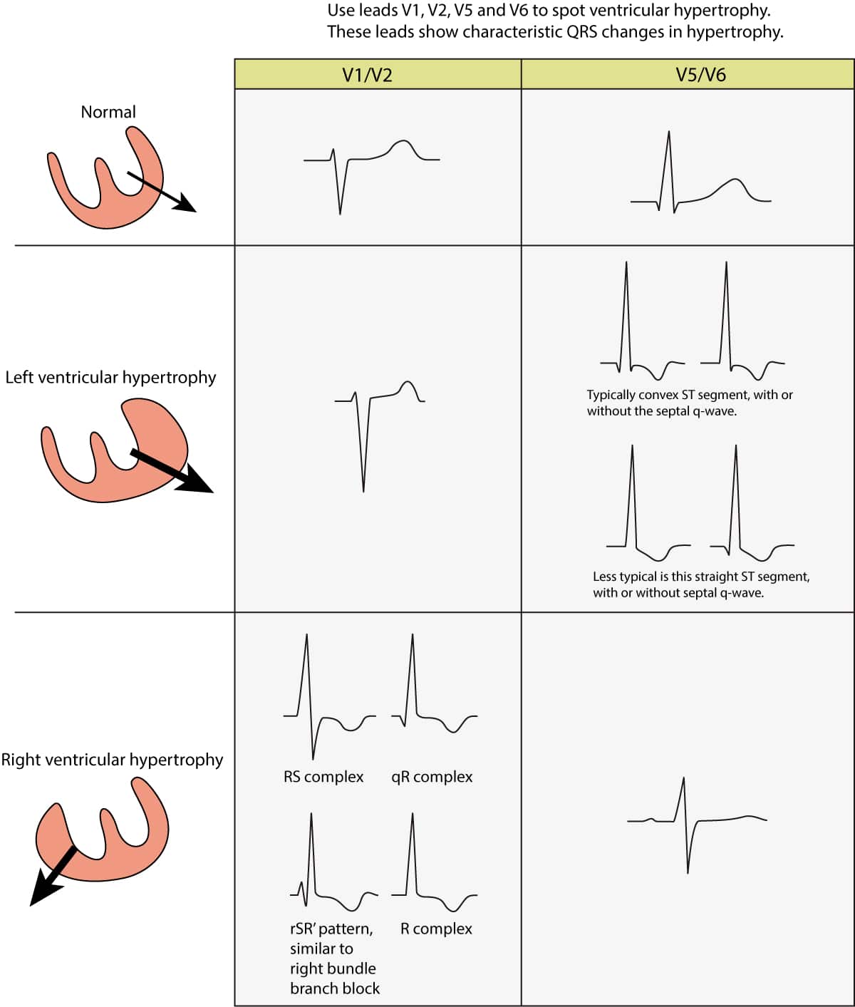
Strain Pattern Ecg Definition. Other abnormalities caused by RVH. Right ventricular strain pattern ST depression T wave inversion in the right precordial V1-4 and inferior II III aVF leads. QRS amplitude voltage criteria. Strain pattern is usually observed in patients with left ventricular hypertrophy andor dilation such as in systemic hypertension or aortic valve disease.

Strain is defined as shortening or lengthening of myocardium. More recent studies utilizing cardiac MRI have examined ECG strain in relation to myocardial tissue changes in patients with aortic stenosis AS. One of the possible causes is poor blood supply to the heart and this is the cause that is most concerning in a person that is about to have elective surgery. Anterior and Posterior Fascicular Blocks. Shortening occurs when myocardium contracts and lengthening occurs when myocardium relaxes stretches out. LV strain pattern with ST depression and T-wave inversions in I aVL and V5-6.
However all patients with left ventricular hypertrophy do not show strain pattern.
ST elevation in V1-3. Anterior and Posterior Fascicular Blocks. Accompanied by ST-segment T-wave changes negativity of ST segment T wave as well as QRS complex changes. Anthony Kashou Left ventricular strain pattern Definition. Right ventricular strain pattern ST depression T wave inversion in the right precordial V1-4 and inferior II III aVF leads. One of the possible causes is poor blood supply to the heart and this is the cause that is most concerning in a person that is about to have elective surgery.